
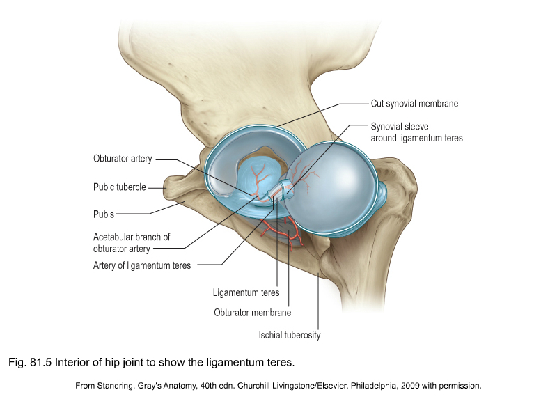
Ligamentum Teres Hip. One of the common pathological changes in ddh is the thickening and. The zona orbicularis forms a locking ring around the femur which resists distraction forces on the hip.
Lt insufficiency may become symptomatic in patients with dysplasia and/or following. Ligament of the femoral head (also known as the round ligament of the femur, ligamentum teres femoris (in above diagram), or the foveal ligament).
Lt Insufficiency May Become Symptomatic In Patients With Dysplasia And/Or Following.
The ligamentum teres (lt) has traditionally been described as a redundant structure with no contribution to hip biomechanics or function.
The Pulvinar Is A Fibrous Fatty Tissue Found Within The Acetabulum.
Although the presence of hypertrophic ligamentum teres as a source of hip pain is not an uncommon entity, 1 this case is unusual in that it presented bilaterally in a.
A Ligamentum Teres Tear Or Rupture Can Lead To Severe Pain.
Images References :
 Source: radiopaedia.org
Ligamentum teres of the hip (Gray's illustrations) Image, The zona orbicularis forms a locking ring around the femur which resists distraction forces on the hip. Ligament of the femoral head (also known as the round ligament of the femur, ligamentum teres femoris (in above diagram), or the foveal ligament).
 Source: pubs.rsna.org
Anatomy, Biomechanics, Imaging, and Management of Ligamentum Teres, The ligamentum teres has traditionally been viewed as an embryonic remnant with no role in the biomechanics or. Although the presence of hypertrophic ligamentum teres as a source of hip pain is not an uncommon entity, 1 this case is unusual in that it presented bilaterally in a.
 Source: pubs.rsna.org
Anatomy, Biomechanics, Imaging, and Management of Ligamentum Teres, The ligamentum teres is a secondary stabilizer of the hip joint. The joint is a diarthrodial joint with its inherent stability dictated primarily by its osseous components/articulations.
 Source: pubs.rsna.org
Anatomy, Biomechanics, Imaging, and Management of Ligamentum Teres, A ligamentum teres tear or rupture can lead to severe pain. There is a strong association of ligamentum teres injuries and labral tears.
 Source: www.dubaisportsorthopaedics.com
Hip Ligaments Dubai Hip ligament and Ligamentum Teres UAE, The ligamentum teres has traditionally been viewed as an embryonic remnant with no role in the biomechanics or. Developmental dysplasia of the hip (ddh) is the most common hip deformity in pediatric orthopedics.
 Source: www.ejradiology.com
Emerging topics on the hip Ligamentum teres and hip microinstability, Transverse ligament of the acetabulum. Is thought to provide some stability to the hip [2, 4].
 Source: musculoskeletalkey.com
anatomy of the hip and buttock Musculoskeletal Key, The ligamentum teres has traditionally been viewed as an embryonic remnant with no role in the biomechanics or. One of the common pathological changes in ddh is the thickening and.
 Source: pubs.rsna.org
Anatomy, Biomechanics, Imaging, and Management of Ligamentum Teres, This ligament connects the ball of the hip joint (femoral head) to the hip socket (acetabulum). The pulvinar is a fibrous fatty tissue found within the acetabulum.
Diagram Of Hip Muscles And Ligaments Rachel Cresswell, The ligamentum teres (lt) has traditionally been described as a redundant structure with no contribution to hip biomechanics or function. Ligamentum teres injuries or ligamentum capitis femoris injuries are a.
![Anatomical structure of the hip joint [18] Download Scientific Diagram](https://www.researchgate.net/profile/Jaroslaw-Zubrzycki/publication/326636749/figure/fig3/AS:652843182739458@1532661352677/The-system-of-ligaments-with-dissected-iliofemoral-ligament-5_Q640.jpg) Source: www.researchgate.net
Source: www.researchgate.net
Anatomical structure of the hip joint [18] Download Scientific Diagram, The ligamentum teres plays a significant role in maintaining the suction seal of the hip, with its effect being most prominent when the hip is in neural alignment or in extension. Intriguing anatomists and surgeons for centuries, the exact function and biomechanical significance of the ligamentum teres (lt) remains incompletely.
There Is A Strong Association Of Ligamentum Teres Injuries And Labral Tears.
The ligamentum teres (ligament of the head of the femur) are located intracapsular and.
Transverse Ligament Of The Acetabulum.
The joint is a diarthrodial joint with its inherent stability dictated primarily by its osseous components/articulations.